
Cervix uteri
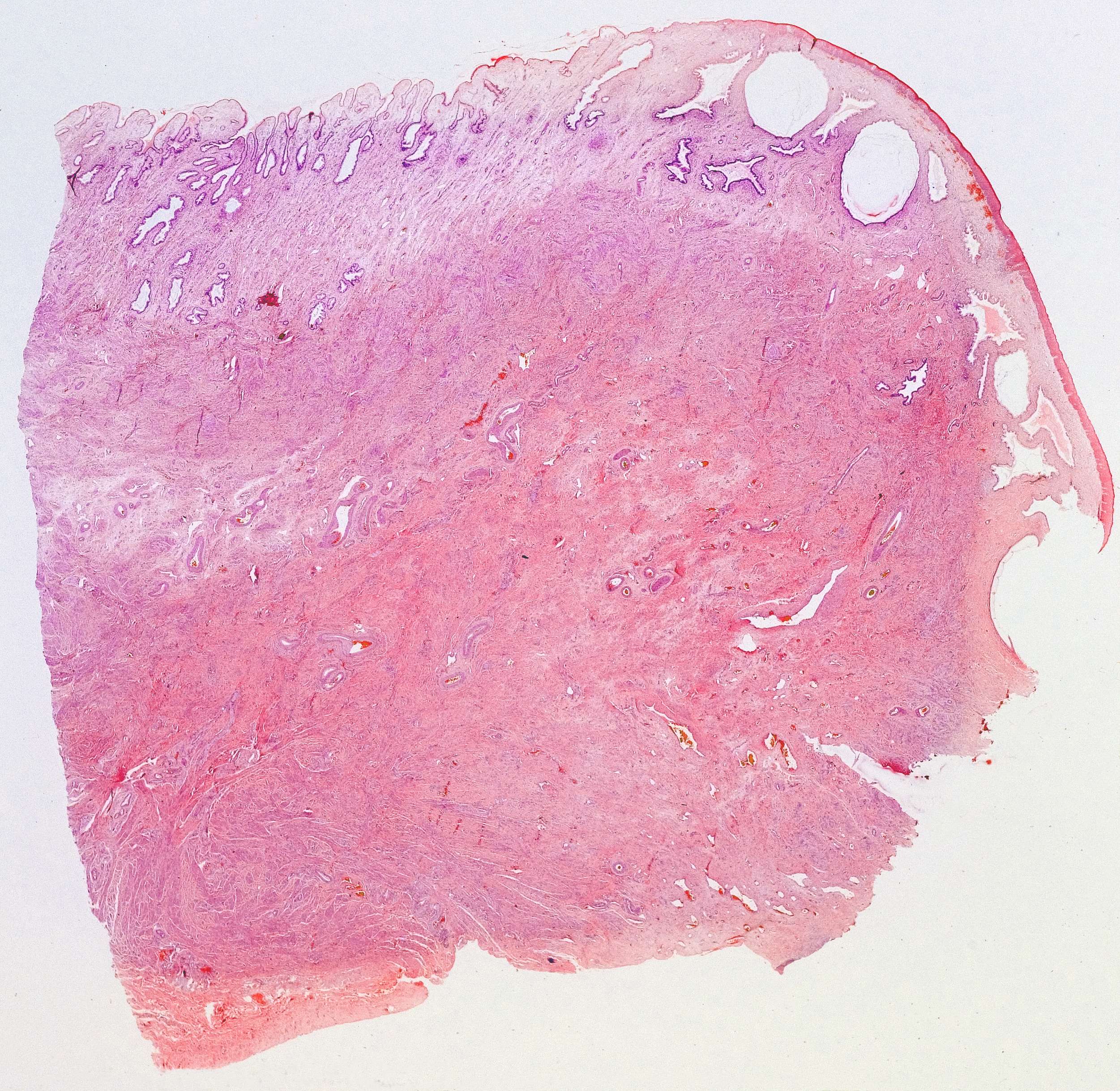
This is an overview of a part of the cervix uteri (the section is cut parallel to the long axis of the cervix). The upper surface of the section (forming the wall of the cervical canal) is covered by the cervical mucosa, and the deeper part, which is more pinkish of colour, is the fibromuscular part of the cervix. It consists mainly of a dense connective tissue with only relatively sparse smooth muscle (compared with the myometrium). The big, white circles are retention cysts (nabothian cysts) of no functional or clinical significance. You see that the wall of the cervical canal is folded. This part is called the endocervix. Further to the right, the surface becomes smooth as it curves downward, covering the portio vaginalis (cervicis). This part is called the exocervix. The next pictures with higher magnification will show details of the different parts. Hit the 'Magnify' button to see these images.