
Penis
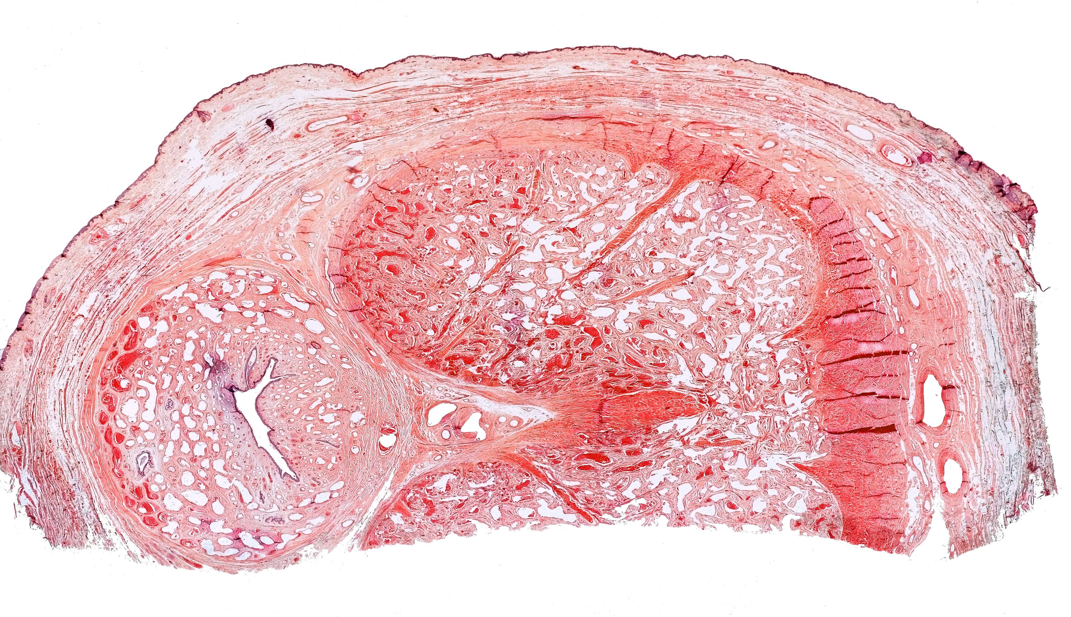
This is a photomicrograph of half the cross-section of the penis. The folded lumen visible at the left side, is the urethra, which is surrounded by the corpus spongiosum. Further to the right you see the corpus cavernosum, covered by a thick tunica albuginea, which also continues into a septum partly dividing the corpora cavernosa. A.dorsalis penis can be seen to the right. The small white spaces in the corpora are caverns, from which the blood has been removed during preparation.
The tunica Dartos, consisting mainly of thin bundles of smooth muscle cells, is visible between the corpora and the epidermis.