
Central veins in pigs liver (100X)
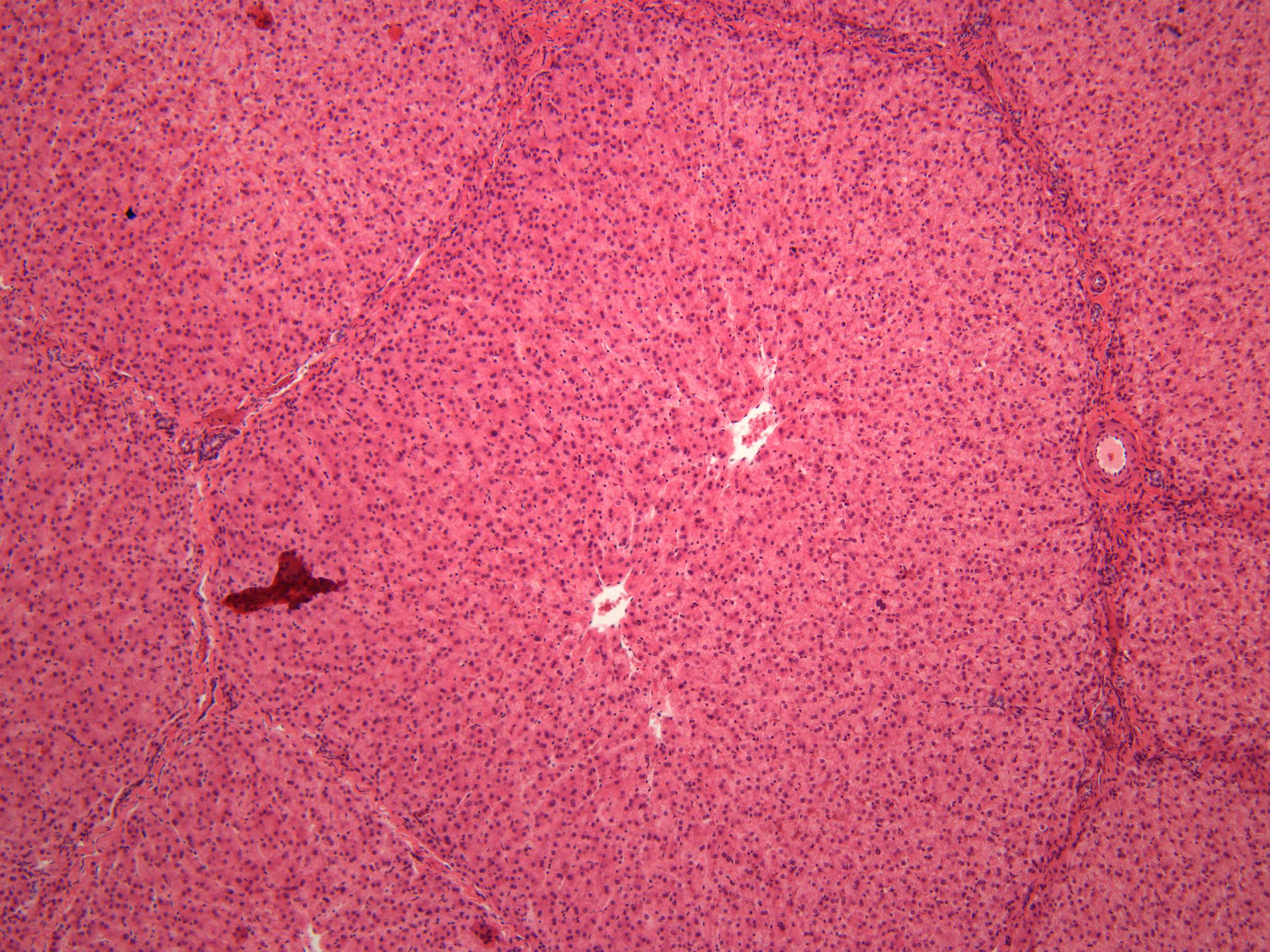
The structural unit in the liver is called a liver lobulus. It is a hexagonal prism that is about 2 mm high and 1 mm wide. A lobulus is bounded by interlobular connective tissue. In a pure cross-section, the lobulus is approximately hexagonal. Often the incision strikes at an angle of the liver lobulus, and then one can see other geometric shapes (triangular, square).
A portal triad is also seen. It consists of these five structures:
- proper hepatic artery, an arteriole branch of the hepatic artery that supplies oxygen
- hepatic portal vein, a venule branch of the portal vein, with blood rich in nutrients but low in oxygen
- one or two small bile ductules of cuboidal epithelium, branches of the bile conducting system.
- lymphatic vessels
- branch of the vagus nerve
The misnomer "portal triad" traditionally has included only the first three structures, and was named before lymphatic vessels were discovered in the structure.