
Tuba uterina (40X)
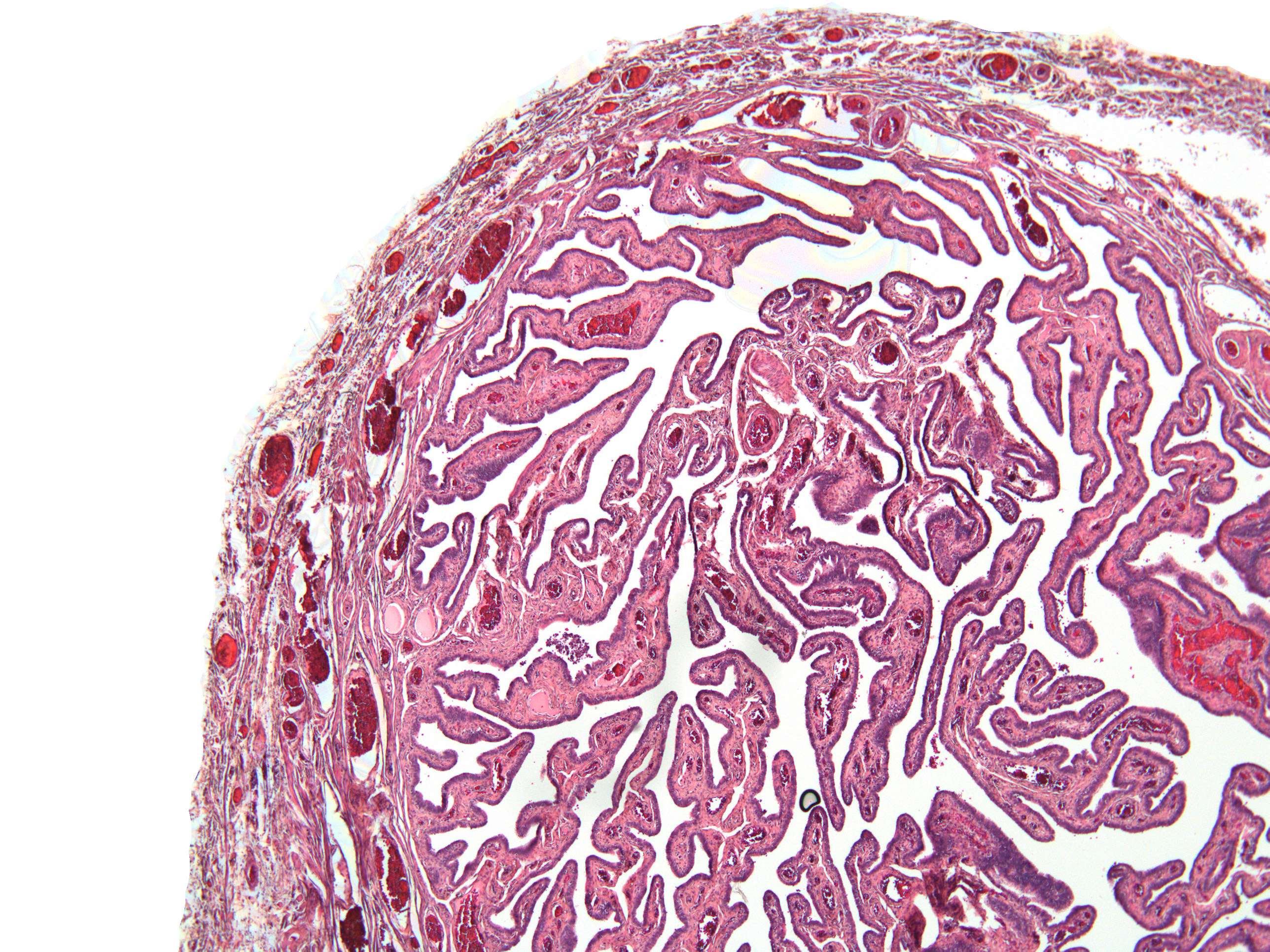
This image display the folds inside the lumen of the tube in some more detail. The pink colour within the folds is the lamina propria, while the thin epithelial covering is seen as a darker line. The wall of the tube contians a muscular layer (tunica muscularis) and one layer of peritoneum. It is not easy to distinguish these layers at this low magnification.