
Ovary (cat) (400X)
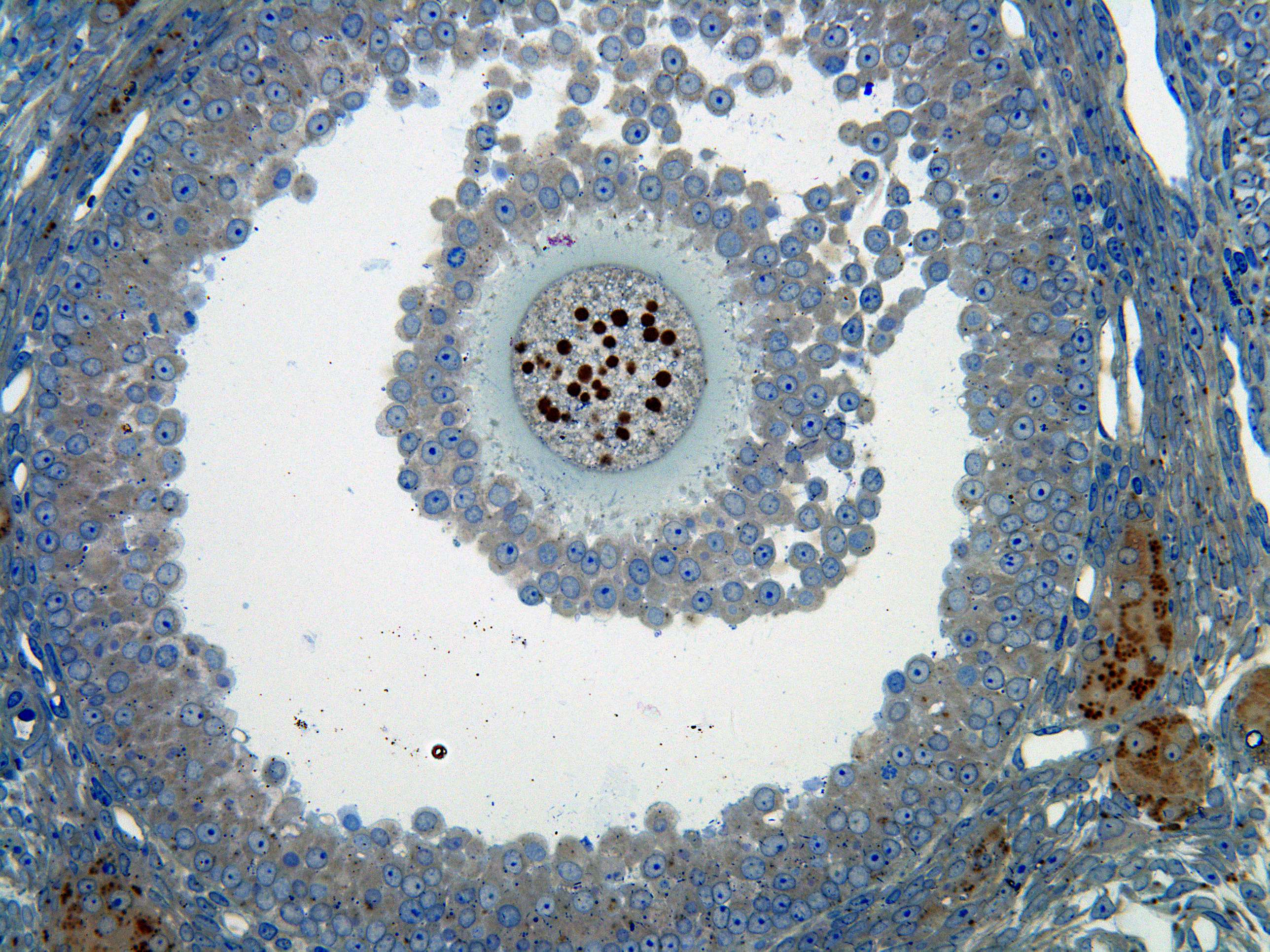
This detailed image displays a secondary follicle with an oocyte, zona pellucida, the granulosa cells, the antrum folliculi, basal membrane and the theca folliculi (interna). Note that there are vessels in the theca interna, but not inside the follicle (a basement membrane separates the granulosa cells from the theca). The brown spots, the lipid droplets, are concentrated in the interstitial cells, the theca cells, and in the oocyte. The lipid contain steroid hormones and their precursors.