
Appendix (100X)
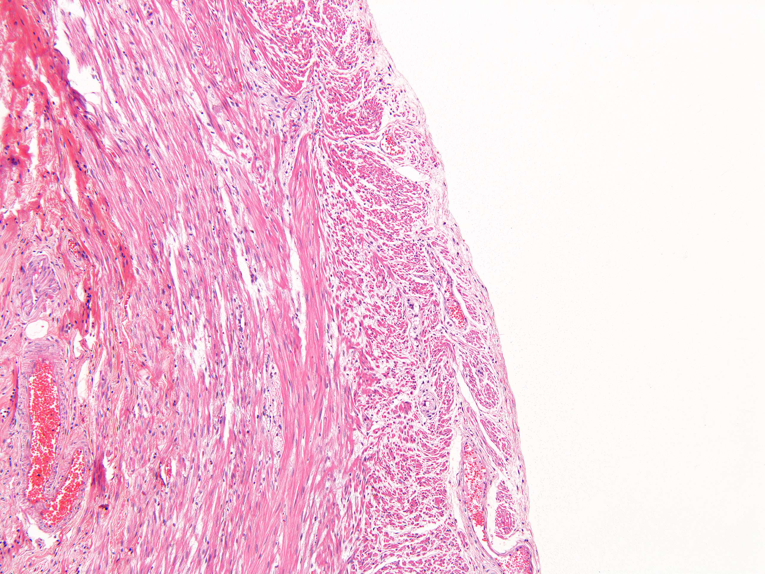
This cross section og the tunica muscularis shows the two main layers beautifully:
- the longitudinal layer most peripherally (fibers cut across)
- the circular layer (fibers cut perpendicularly)
Further inwards you can see the submucosa. The outer surface of the appendix facing the abdominal cavity is covered by the tunica serosa.