
Appendix (400X)
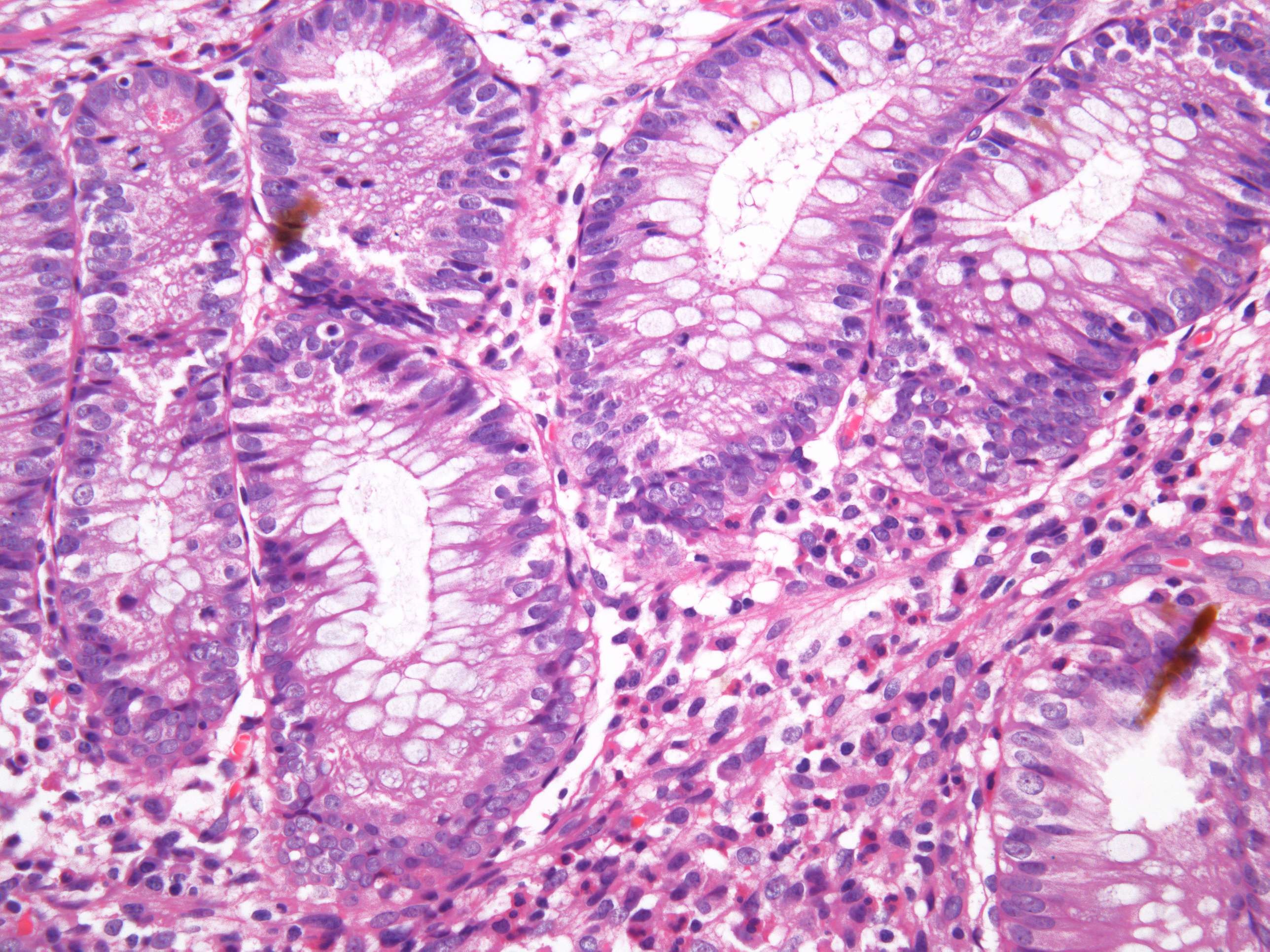
This image show som distinct feutures of the crypts. You can see the lumen of the crypt surrounded by the apical part of the columnar epithelial cells. The nuclei of the columar cells are seen basally in the cells almost forming a magenta band of small dots lining the outermost part of the crypt next to the basal lamina. The lamina propria surrounds the crypts.