

Chronic hyperplastic pulpitis (pulp polyp) (40X)
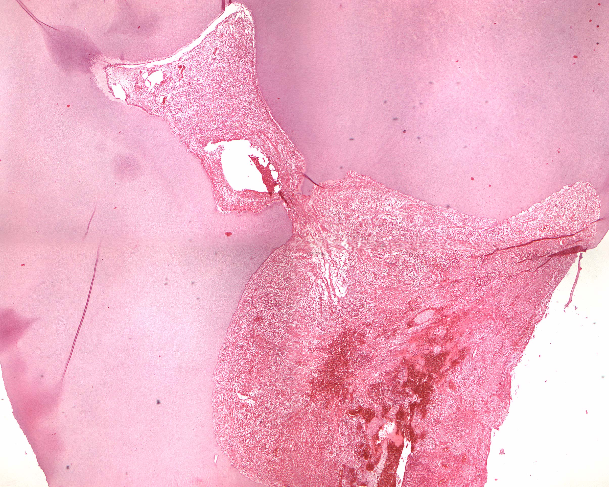
Clinical information: A reddish tissue masses protruded from a carious lesion in tooth 46 in a 10-year-old patient protruded. The tooth was extracted.
Microscopic examination: The image shows a molar with a large cavity and a small opening to the pulp (caries ad pulpam). Pronounced inflammatory changes are found within the pulp, which is penetrated by leukocytes and partially transformed into granulation tissue. Remains of odontoblasts are seen against the dentin and a small, empty cavity compatible with a pustule (small, pus-filled cavity) in the pulp. Granulation tissue protrudes through the opening of the pulp and fills the cavity (pulp polyp; polyp from G, polys=many, pous=foot; a broad-based or stalked, smooth protuberance). In the crown pulp chamber, in addition to chronic inflammatory infiltrate and dilated veins, a number of larger, free denticles are seen. Towards the apex, diffuse calcifications (basophilic) appear arranged parallel to the direction of the veins.