

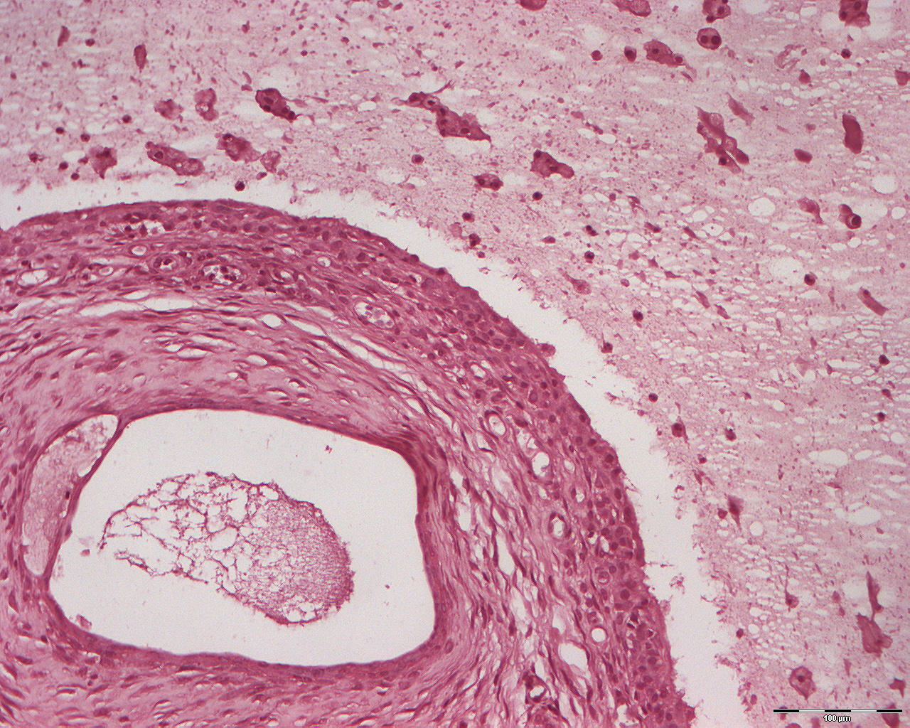
Oral mucocele (extravasation mucocele) and chronic sialadenitis (200X)
Clinical information: A 32-year-old woman presented herself with a swelling on her lower lip that she had had for a long time.
Clinical diagnosis: Mucocele (mucus(L) = mucus; kele(G) = swelling, tumor).
Microscopic examination: The typical appearence of an extravasation mucocele is seen. There is a larger cavity which is limited by a sort of rim that is rich in fibroblasts and macrophages and filled with an eosinophilic mass (mucus) with a good number of leukocytes and tissue herniation. In close proximity to the lumen, salivary gland tissue with parenchymal loss, fibrosis, chronic inflammatory cells and basophilic substance (congestion of mucus) is seen in well dilated excretory ducts. The mucocele lacks an epithelial lining and is therefore not a cyst.
Comment: The chronic inflammation seen in the salivary gland tissue poses a risk of relapse if the inflamed glands are not extirpated.