

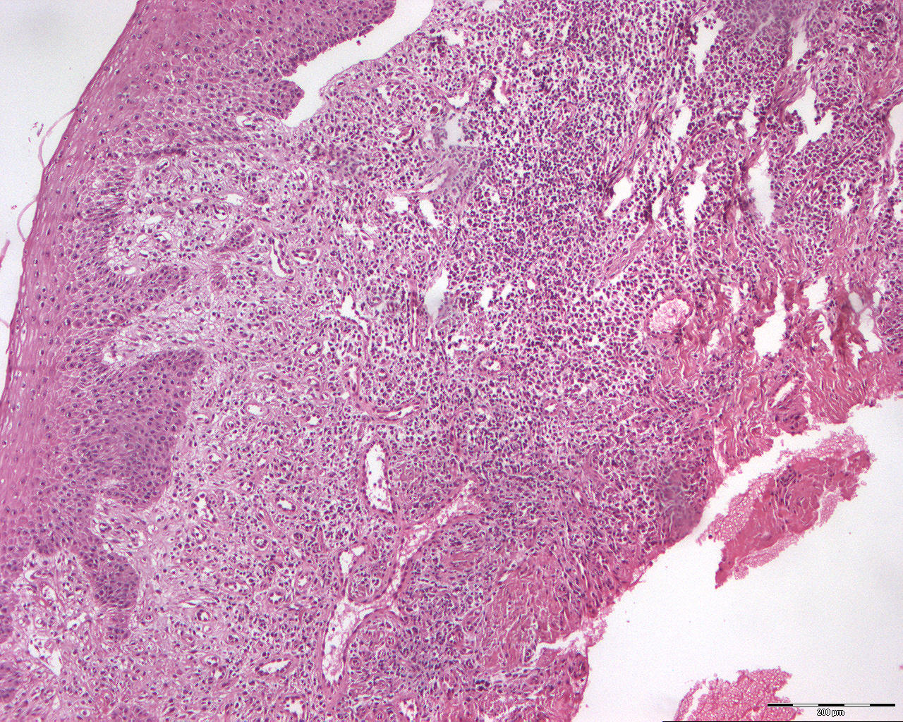
Mucous membrane pemphigoid (100X)
Clinical information: A 73-year-old woman had a erythematous, slightly edematous looking gingiva in the front of the upper jaw. When touched, the mucosal surface loosened (so-called desquamation). The gingiva was not painful but somewhat unpleasant.
Clinical diagnosis: Mucous membrane pemphigoid (Greek: pemphix = bladder; eidos = shape).
Microscopic examination: Microscopically, the mucosa is covered with parakeratinized and non-keratinized multi-layered squamous epithelium, which forms short ridges against the underlying connective tissue and is in some places demarcated in a straight line from this (atrophic). Furthermore, a subbasal slit is seen (top of image). The underlying connective tissue shows a moderate to intense infiltration of lymphocytes and plasma cells with diffuse distribution in the deeper part of he tissue.
Comment: In contrast to lichen planus, no primary basal cell degeneration takes place in benign mucosal pemphigoid or pemphigoid. "Sawtooth" proliferation of the epithelium is also never found, and the inflammatory infiltrate has a diffuse distribution in the deeper parts and shows a heterogeneous cell population with lymphocytes, plasma cells and sometimes islets of granulocytes. Immunologically, one will find a band-shaped drop of immunoglobulins, preferably IgG and complement factor 3 (C3) at a subbasal level. Such a reaction does not occur with lichen planus.