

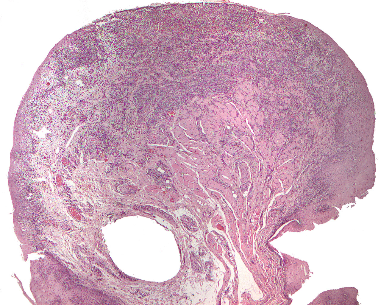
Aphthous stomatitis (40X)
Clinical information: A 20-year-old man had 2 weeks previously had petechiae-like lesion covered by a yellowish coating in the buccal fold of the lower jaw as well as the buccal mucosa. The changes regressed, but after a week multiple, aphthous-like lesions with a diameter of 14 mm appeared in the buccal mucosa, associated with a strong burning sensation. The patient had submandibular lymphadenopathy but the lesions did not affect his general condition.
Clinical diagnosis: Multiple aphthous ulcerations. The preparation is taken from the canine mucosa at the reion of tooth 44.
Microscopic examination: Microscopically, small mucosal pieces of tissue can be seen whose surface on both sides is covered by nonkeratinized to parakeratinized, regular and well-defined multi-layered squamous epithelium with significant spongiosis and the presence of large, light-colored stratum spinosum cells with a small and dark nucleus. The epithelium proliferates to a limited extent in depth, forming somewhat spikes of variable length. The subepithelial loose connective tissue is diffusely infiltrated, mildly to moderately, by lymphocytes and plasma cells as well as some granulocytes in perivascular localization. Deeper, one finds some striated musculature with moderate infiltration of lymphocytes and plasma cells between the muscle bundles. In some places, the surface is ulcerated and covered by fibrin deposits with accumulated cellular elements. In the underlying granulation tissue there is intense infiltration of lymphocytes, plasma cells and granulocytes as well as some hyperemic capillaries.
Comment: The histological picture of aphthous stomatitis is non-specific, but the available material is compatible with this condition (aphta, Greek = small wound, from aptein to set fire to).