

Oral lichen planus (40X)
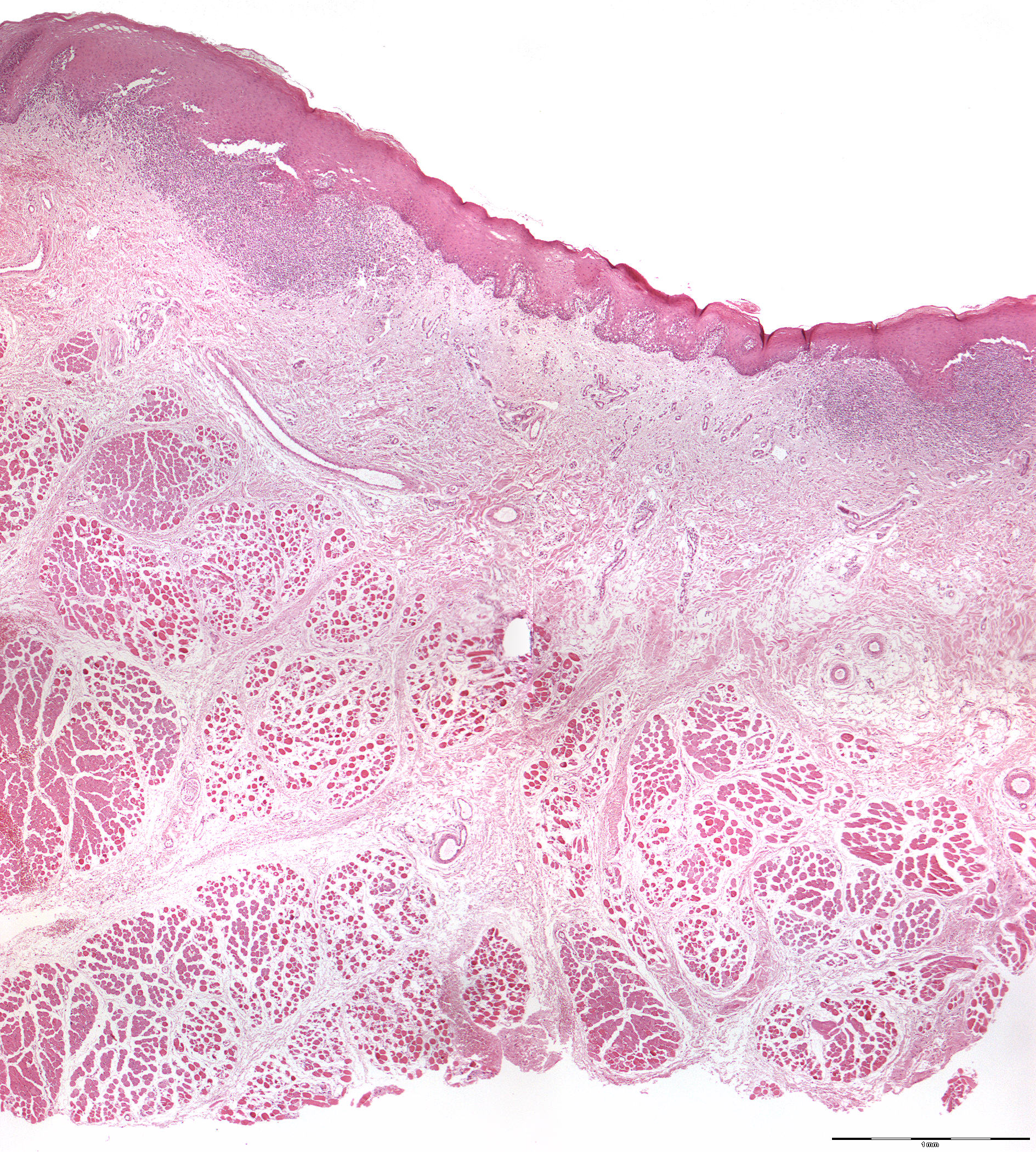
Clinical information: A 10-year-old boy had allegedly for a few months had a now approx. 10 mm large whitish, non-painful lesion with slightly indurated lines on the mucosa of the right cheek. The entire lesion was extirpated with approx. 3 mm margin against surrounding healthy tissue.
Clinical diagnosis: Lichen planus (Latin: lichen = low botanical; planus = flat).
Microscopic examination: Microscopically, the mucosa is covered by hyperparakeratinized multi-layered squamous epithelium, which is relatively thick with wide extensions towards the underliing tissue. Elsewhere, the epithelium is thin and without spikes (atrophic), in other places it forms short, pointed spikes against the underliing tissue. Especially in the atrophic areas, one finds some vacuolization (formation of vacuoles) of the basal cell layer and some lymphocytes in the deep epithelial layer, as well as an eosinophilic coagulum close to the border with the connective tissue. Furthermore, an irregular gap is seen, partly subbasal and partly deeper within the connective tissue. This is an artefact that is often seen with lichen planus and which shows poor tissue cohesion in the area in question. Furthermore, a superficial, almost ribbon-like dense lymphocyte infiltrate is found. Deeper, one finds inconspicuous loose connective tissue, fatty tissue and striated muscle towards the resection margin.
Comment: Immunofluorescence microscopy shows no deposition of immunoglobulins or C3 in the basement membrane zone of lichen planus, but only a shaggy band of fibrinoid. This corresponds to the eosinophilic coagulum in H+E stained preparations.