

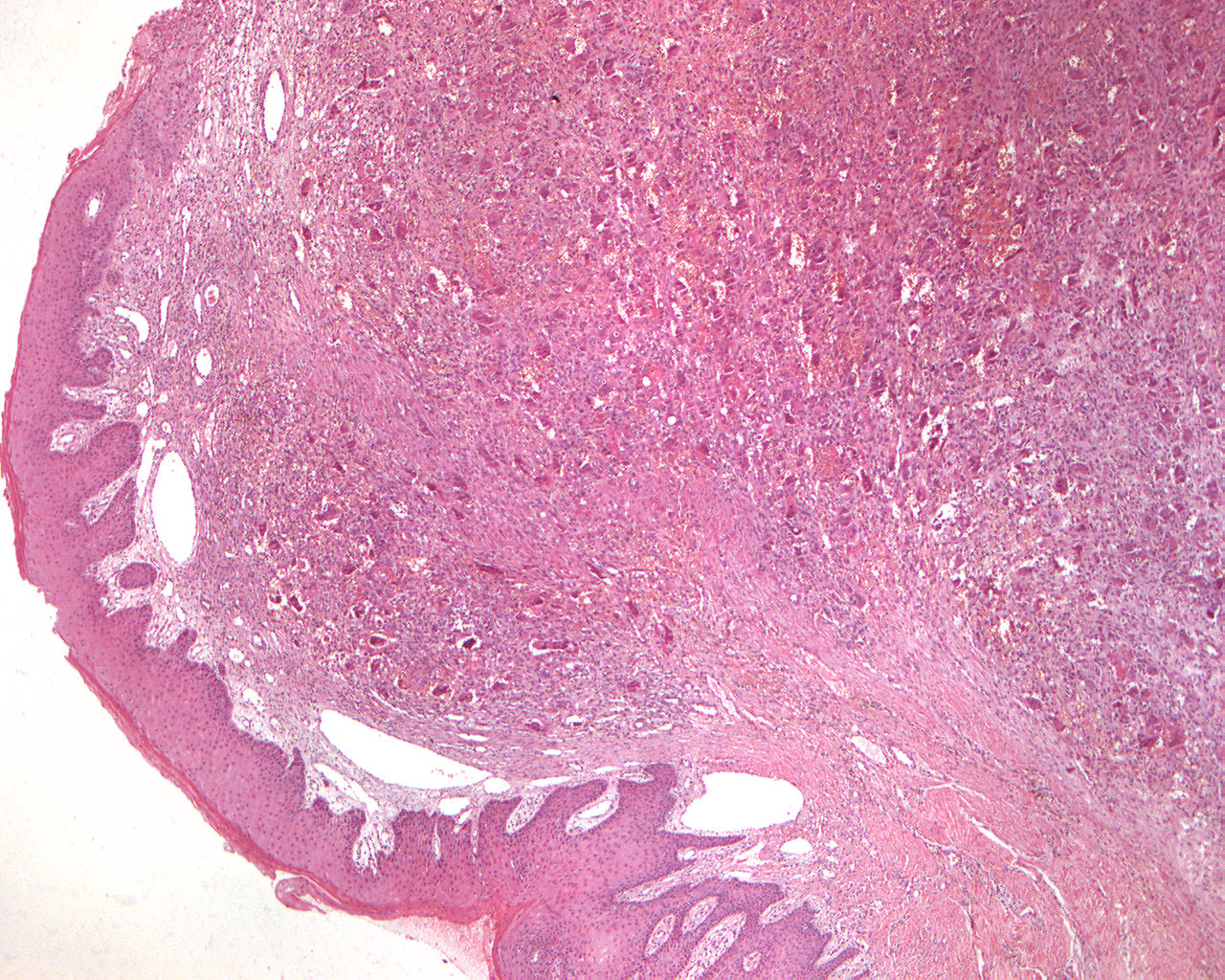
Peripheral giant cell granuloma histology (40X)
Clinical information: A 62-year-old man had noted a painless "dental abscess" in close to the tooth 35 which had grown and decreased in size three times over the past month. The use of a partial denture resulted in some soreness. The tumor presented as a blue-colored, fibrin-covered, well-demarcated , broad-stemmed and bulging tumor of about 1.5 cm in diameter.
Clinical diagnosis: Tumor gingivae.
X-ray findings: Irregular bone crest.
Microscopic examination: The specimen was a broad-based pieces of mucous membrane with a strongly prominent surface which are partially covered on both sides by hyperparakeratinized, regular, somewhat hyperplastic multi-layered squamous epithelium with a sharp demarcation from the underlying connective tissue. Above the highest prominence of the bulge, an ulceration with fibrin deposits and some other cellular elements is seen. Subepithelially, a narrow zone of chronically inflamed connective tissue is found. The main component of the prominence is made up of somewhat deeper connective tissue rich in large fibroblasts and with an impressive amount of giant cells of the foreign body type. These are often found in vein walls and intravascularly. Furthermore, the picture is characterized by numerous small, thin-walled veins and vein clefts, and scattered occurrences of mononuclear inflammatory cells are found. The special granulation tissue extends all the way to the resection margin in depth. What other pathological conditions have the same histological picture?