

Actinomycosis of the mucous membrane (40X)
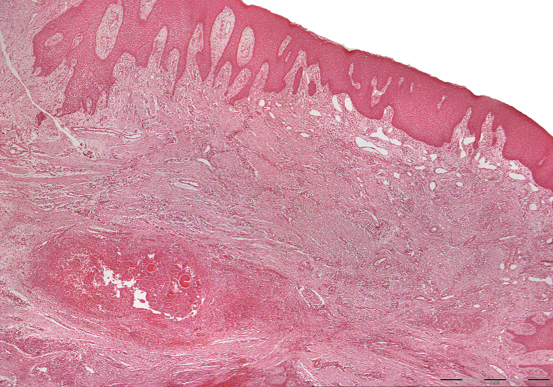
Clinical information: A 56-year-old man with a complete upper jaw prosthesis had increasing pain in the edentulous region of 21, 22 and 23. A boat-shaped piece of mucosa was removed from the area in question.
Clinical diagnosis: Infected cyst of the upper jaw.
Microscopic examination: The mucosa is covered by multi-layered squamous epithelium with a parakeratinized surface (parakeratinized: an epithelium that is characterized by incomplete keratinization of the cells in the stratum corneum). The stratum spinosum is somewhat increased in thickness, and the epithelium proliferates in places in a net-like manner and somewhat cone-shaped in det deeper parts. The basal layer is distinct and well demarcated from the underlying loose connective tissue, which is somewhat oedematous and relatively rich in small, dilated, partly blood-filled veins. One finds moderate to intense infiltration of inflammatory cells, both mononuclear and polymorphonuclear. Deeper, somewhat more fibrous connective tissue is seen, which partly surrounds the pustules. Here, in addition to cellular elements, several irregular, lobular, basophilic structures with an eosinophilic periphery with a diameter of approx. 0.1 mm is seen. The structures are fairly homogeneous centrally but exhibit radiating filaments in the periphery. The image fits with Actinomyces colonies, which was confirmed in PAS-stained material. Actinomyces colonies can be observed in plaster with the naked eye as so-called sulfur grains (yellow colour).