

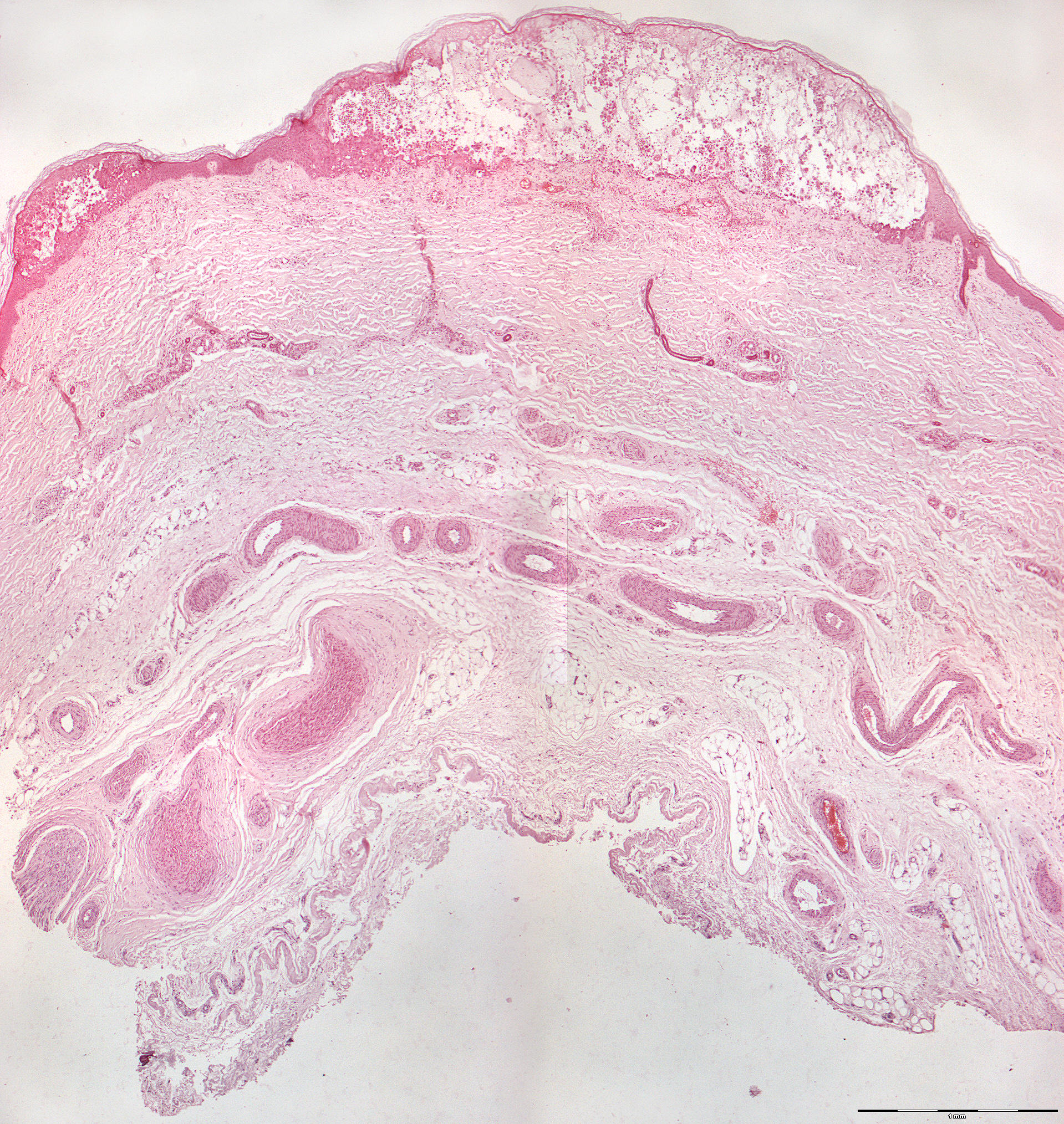
Herpes zoster histology (40X)
Clinical information: A 66-year-old man was treated with radiation and cytostatics as well as painkillers for metastatic bronchial carcinoma. The patient died severely emaciated and during the last days of his life had been troubled by a skin rash with groups of blisters on the lower limbs, in the groin and on the thorax. The section is from the autopsy.
Clinical diagnosis: Herpes zoster generalisata (Greek: herpein = to crawl: zoster = belt).
Microscopic examination: The speciman displays pieces of skin with blisters where the roof of the blisters consists of relatively well-preserved multi-layered squamous epithelium. Deeper in the tissue, only remnants of the basal cell layer are visible. The lumen of the blister itself is permeated by a network of epithelial cell remnants. This reticular degeneration occurs when the epithelial cells are distended by intracellular edema so that the cell walls rupture. Adjacent cells fuse together, and a multilocular vesicle is formed, where septa form at the remaining and more resilient cell walls. A number of large, bloated cells with eosinophilic cytoplasm with one or more nuclei are also seen scattered throughout. These "balloon cells" are degenerated, virus-infected epithelial cells (ballooning degeneration). In some places, eosinophilic inclusion bodies can be found in the nuclei themselves (intranuclear inclusion bodies). These inclusion bodies represent sites where viral replication takes place/has taken place.
The intercellular connection between the balloon cells has been dissolved (acantholysis). The epithelial cells in the basal layer also show degenerative changes with disturbances in the chromatin. On both sides of the walls of the blister, one also finds highly swollen epithelial cells that are characteristic of a viral infection. Furthermore, a number of leukocytes are found in the blister. Mild infiltration of lymphocytes and plasma cells (immunosuppressed patient) and some angiectasias are seen in the subepithelial connective tissue.
The picture is identical to that found in chickenpox (varicellae) and herpes simplex.