

Acute necrotizing ulcerative gingivitis (ANUG) (40X)
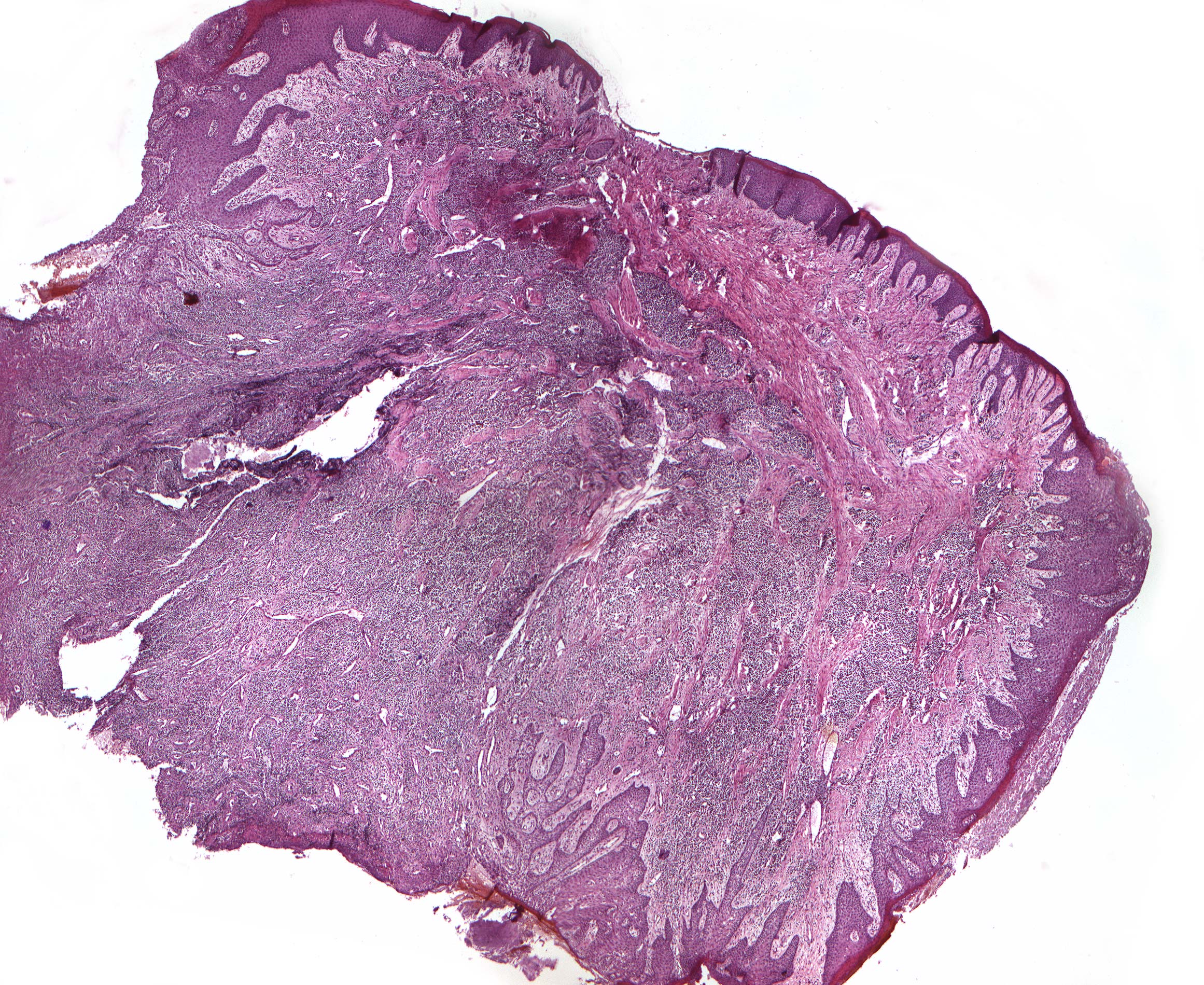
Clinical details: A 20-year-old military recruit had painful, erythematous and edematous interdental papillae with crater-like, greyish depressions at the apex that made the papillae look like they were cut off.
Clinical diagnosis: Acute necrotizing ulcerative gingivitis.
Microscopic examination: Microscopically, the mucosa is seen to be covered with hyperparakeratinized multi-layered squamous epithelium, which proliferates in depth with varying deep ridges and network formation. Enlarged intercellular spaces are seen (intercellular edema = spongiosis). Above the preparation's highest prominence corresponding to the papilla tip, there is a thick fibrin deposit and covered by large accumulations of basophilic substance compatible with plaque. Below the ulceration and subepithelially, granulation tissue with intense infiltration of lymphocytes, plasma cells and some granulocytes is found. Dilated, leukocyte-containing capillaries with somewhat thickened walls are also seen here.