

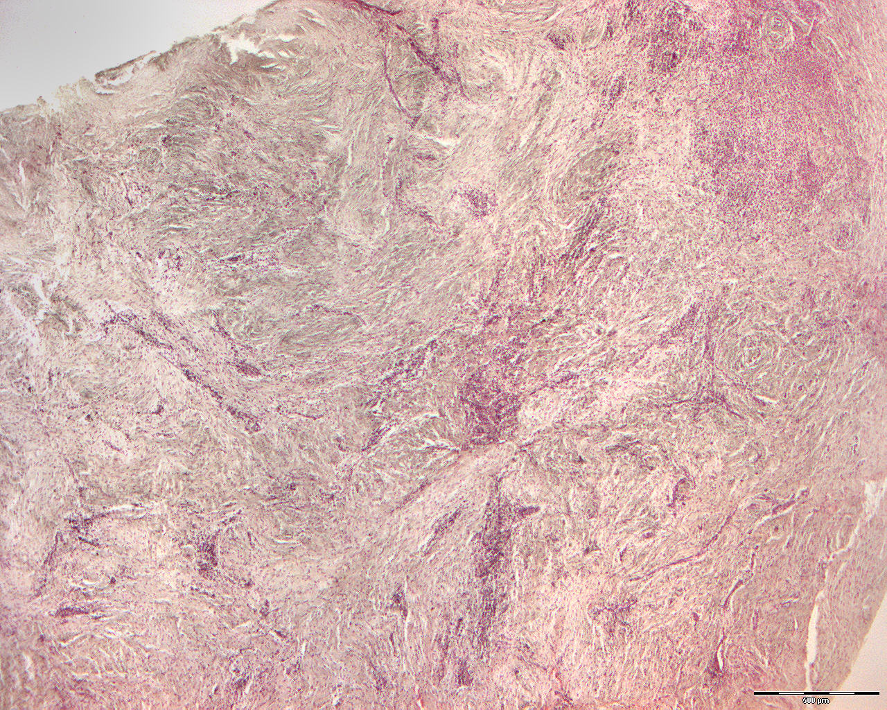
Granuloma from apex of a tooth with chronic inflammation (40X)
Clinical information: A 35-year-old woman had a walnut-sized, cystic radiolucency (transparent to X-rays) apical to teeth 41 and 42. Surgicaly the tissue was removed. Makroscopically it resebled fibrous granulation tissue.
Clinical diagnosis: apical periodontitis teeth 41, 42.
Microscopic examination: Microscopically, oval pieces of soft tissue are seen which are mostly exfoliated by a connective tissue capsule with patchy infiltrates of mononuclear inflammatory cells, mainly lymphocytes and some plasma cells. Centrally, rather low-fibre, relatively cell-rich connective tissue is seen with a significant accumulation of mononuclear inflammatory cells, most of which are lymphocytes but a good number of which are also plasma cells.
Comment: the image is rather dull