

Squamous granuloma from the apex of tooth 22 (40X)
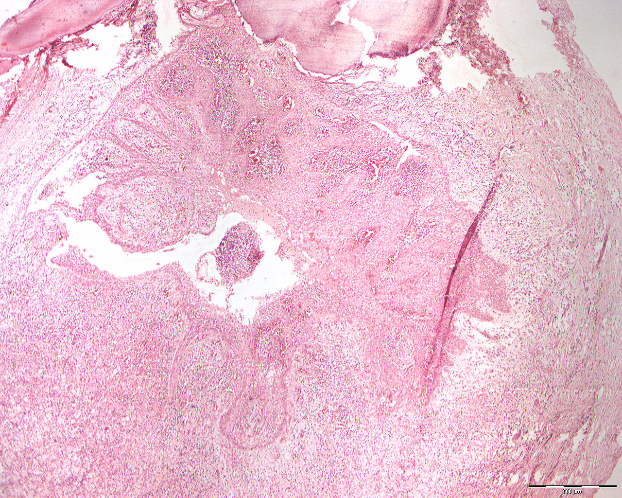
Clinical information: In a 54-year-old man, a routine X-ray examination revealed a small, well-defined radiolucent lesion apical to the tooth 22. An apical amputation of the root was performed.
Clinical diagnosis: Apical granuloma
Microscopic examination: One can see round / oval pieces of tissue that are peripherally limited by a connective tissue capsule and some basophilic, herniated material that represents part of an uncalcified tooth apex. Inside the connective tissue capsule is a layer of cell-rich granulation tissue dominated by lymphocytes, plasma cells and large, bright lipophages (fat-eaters, foam cells). More centrally, an irregular lumen is seen lined by multi-layered squamous epithelium with significant hyperplastic growth in the form of networks and spikes.