

Tuberculous submandibular lymphadenitis (40X)
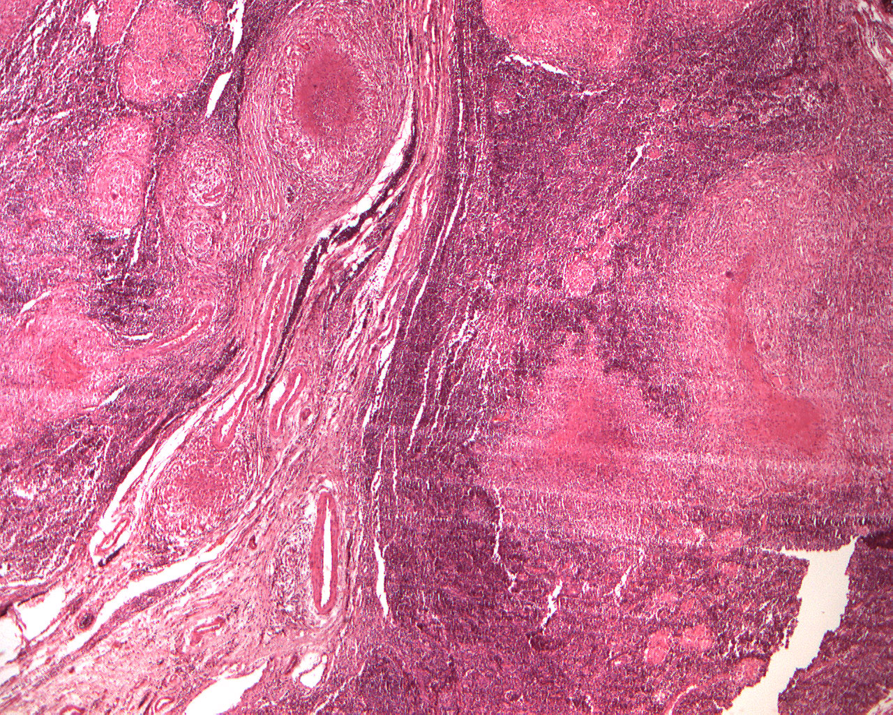
Clinical information: The material has been removed from a tumor in the submandibular region.
Clinical diagnosis: Salivary gland tumor?
Microscopic examination: Lymph nodes are found permeated by granulomas which centrally consist of eosinophilic, necrotic, structureless fine granular masses (caseous necrosis, Latin: caseus=cheese). Peripheral to the necrosis, dense aggregates of relatively large cells with eosinophilic cytoplasm and a relatively large, vesicular nucleus (epithelioid cells = large macrophages) and scattered giant cells with horseshoe-shaped nuclei (Langhan giant cells) are seen. One does not find any of the lymphocyte swelling typical of tuberculosis in this case, as the tissue is lymphatic.