

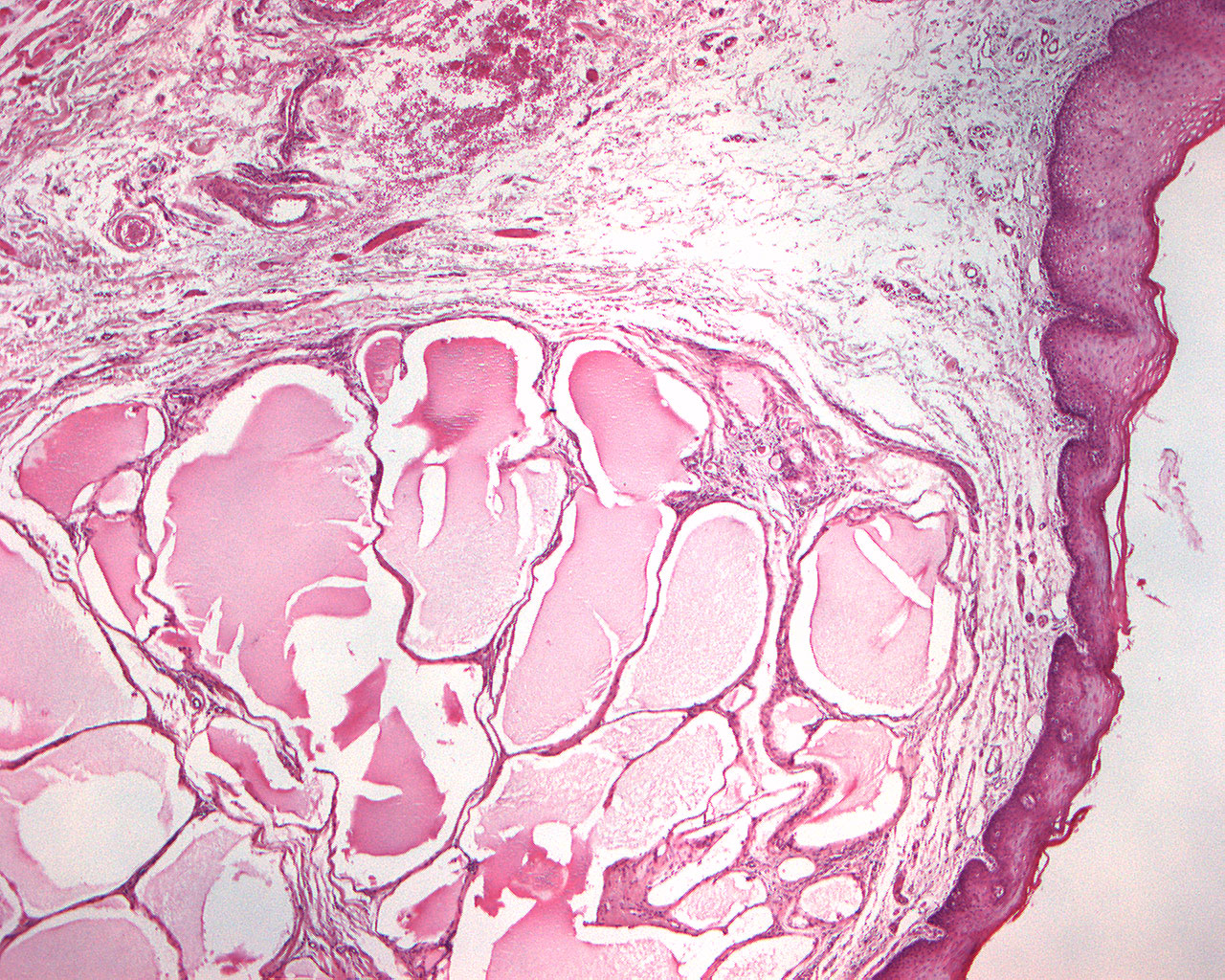
Lymphangioma (40X)
Clinical information: A 53-year-old woman had had a swelling of her right side of her lower lip for 5-6 years. The swelling was often traumatized by biting, otherwise it produced no symptoms.
Clinical diagnosis: Retention cyst.
Microscopic examination: Microscopically, broad-based pieces of mucosa with a slightly convex surface are seen, covered by hyperparakeratinized and partly hyperorthokeratinized, largely narrow, regular, four-layered squamous epithelium with a sharp demarcation against the underlying, loose connective tissue. There are numerous, closely spaced, dilated, irregular, thin-walled veins lined by a single layer of endothelial cells. The lumen is mostly filled by eosinophilic masses (plasma proteins). A thin connective tissue capsule is seen and, in the deeper part of the tissue, some striated musculature is seen.
Comment: A lymphangioma is a rare benign tumor or malformation of the lymphatic system, usually present at birth and commonly found in the head and neck region. There are three main types: capillary lymphangiomas, cavernous lymphangiomas, and cystic hygromas. Treatment depends on the size and symptoms, with options including surgery, laser therapy, sclerotherapy, or medication.