

Schwannoma (40X)
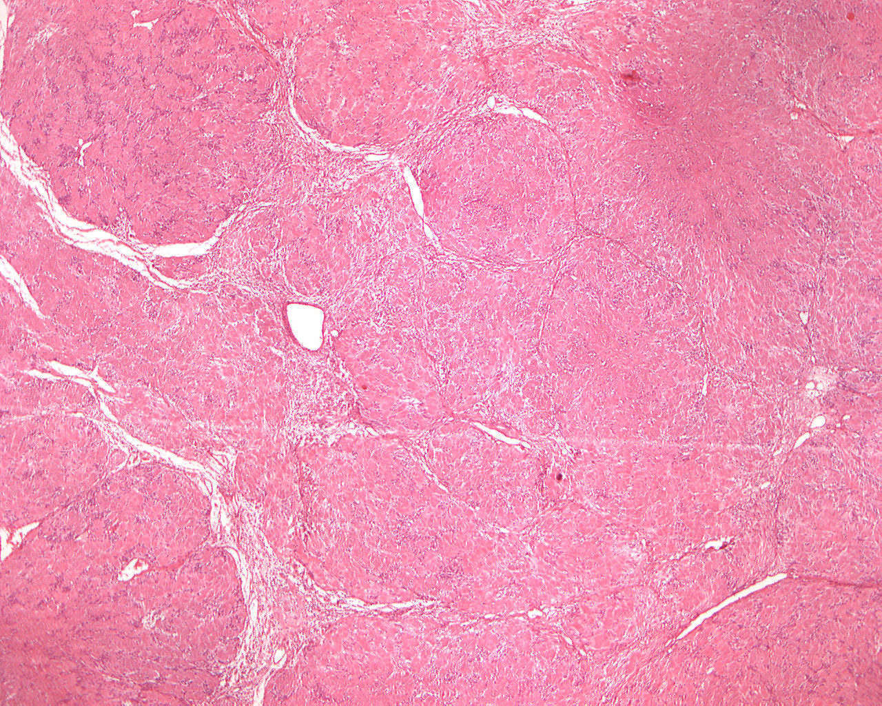
Clinical information: A 20-year-old woman had allegedly had a tumor in the left sublingual region for more than 10 years. The tumor was observed peroperatively to be encapsulated, easy to remove and had a firm consistency.
Clinical diagnosis: Tumor of the sublingual region.
Microscopic examination: Microscopically, the large pieces of tissue are mostly made up of eosinophilic, areas with few nuclei (mostly cytoplasm) with palisade-arranged, round-oval or elongated nuclei peripherally (Antoni type A tissue). Two rows of nuclei with an intervening area containing few nuclei are termed a Verocay body (organoid structure). Scattered within the tissue specimen, small areas are also seen where loosely structured, somewhat edematous looking tissue predominates (Antoni type B tissue). Both tissue types are considered derived from Schwann cells (neural limb).