

Squamous cell carcinoma histology (40X)
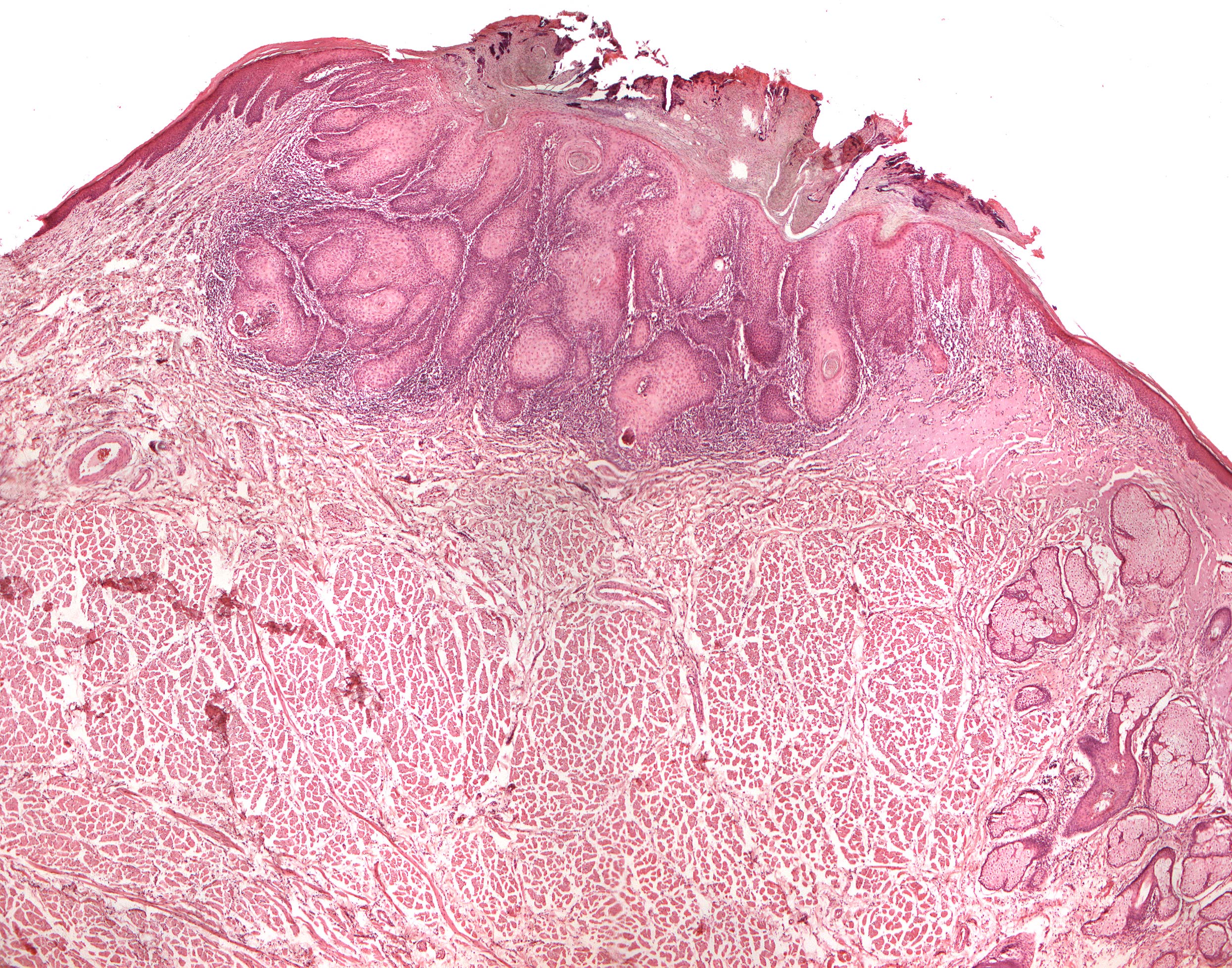
Clinical information: A 60-year-old man had a hard, tumor-like growth on the lower lip, just lateral to the midline. A small sample biopsy (incisional biopsy) showed squamous cell carcinoma. Our section is taken from the following wedge operation.
Clinical diagnosis: Tumor labii
Microscopic examination: The image shows a cross-section of the lip with the skin and its attributes (hair follicles, sebum and sweat glands) on one side, mucous membrane with salivary glands on the other side. Above the highest bulge, an epithelial tumor tissue is seen which grows infiltrating in depth. The surface is significantly keratinized, and horn pearl formation occurs within the stratum spinosum. The basal cell layer is relatively untidy with polymorphic cells and some mitoses. Subepithelial infiltration of lymphocytes and plasma cells is relatively pronounced. Note the sharp transition between normal epithelium and tumor epithelium.