

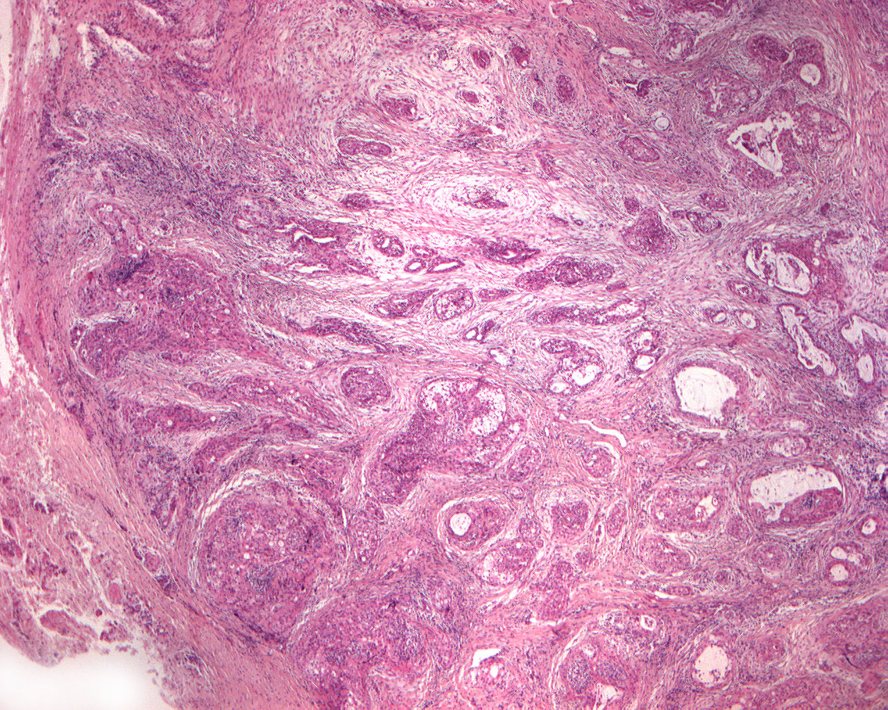
Squamous carcinoma (40X)
Clinical information: A 16-year-old girl was referred to an oral surgeon for a tumor-like lesion in the palate.
Clinical diagnosis: Tumor palati.
Microscopic examination: The tumor tissue, which is partially surrounded by a capsule-like structure, has 2 main components;
- islands and strands of squamous epithelium without striking polymorphism
- mucus-producing cells arranged in gland-like structures and spread out in the squamous epithelial tissue.
The tumor is permeated by tracts of fibrous connective tissue which in places show chronic inflammation.