


Mineralization of the cementum
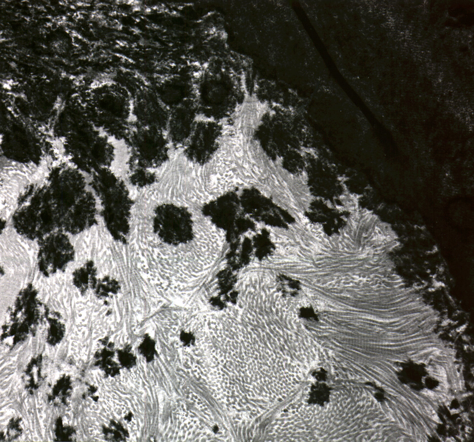
This section shows the mineralization process of the cementum. The "finished cementum" can be seen to the right, the precementum to the left (comprised of collagen fibrills and minreralizing cementum).
The precementum is filled with a matrix of fibrillar collagen. One can see how the apatite crystals has grown within the collagenous matrix of the precementum forming circular areas of mineralized precementum. The image is acquired using transmission electron microscopy (TEM).