


Dentinal tubules viewed from the pulp
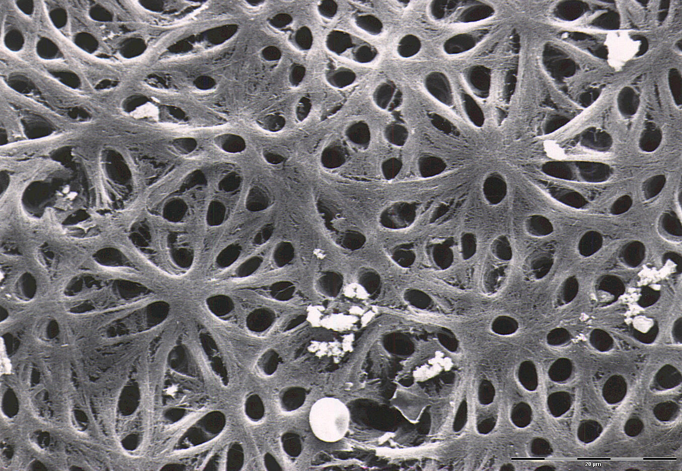
This image shows predentine with dentinal tubules viewed from the pulpal side of the dentin. The odontoblasts have been removed. As in the other electron microscopic images of the dentinal tubules, an erythrocyte has been included the image. This makes it easier to understand the width of the tubules. A healthy erythrocyte is about 7,5 µm in diameter. If you take a look at the images from the dentine close to the dentino-enamel junction (DEJ), you'll see that the dentinal tubules are much wider closer to the pulp than at the DEJ.