

Pleomorphic adenoma histology (40X)
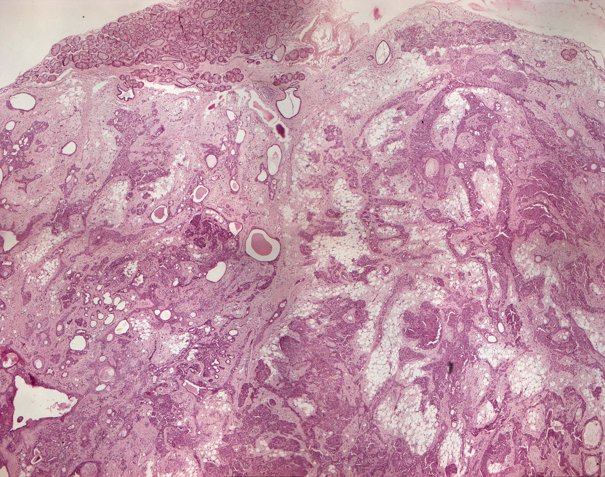
Clinical information: A 62-year-old woman had a tumor-like thickening in the parotid region. The lesion had slowly increased in size over several years.
Clinical diagnosis: Parotid tumor.
Microscopic examination: Benign-looking, epithelial tumor tissue is seen arranged in flakes, islands and strings as well as in cystic and tubular structures. The epithelial cells are of two types; one type is rather poorly distinct, eosinophilic cells that make up most of the tumor tissue, the other type is polygonal cells with clear cytoplasm and angular or oval, dark nuclei (myoepithelial cells/basket cells, which have contractile properties). The epithelial tissue is clearly demarcated from the stromal connective tissue but have a diffuse transition into the mesenchymal tumor component which consists of hyaline, myxoid and chondroid (cartilage-like) tissue. The tumor also contains a good amount of fatty tissue. The tumor is encapsulated, but notice the tumor tissue that lies within the capsule.