


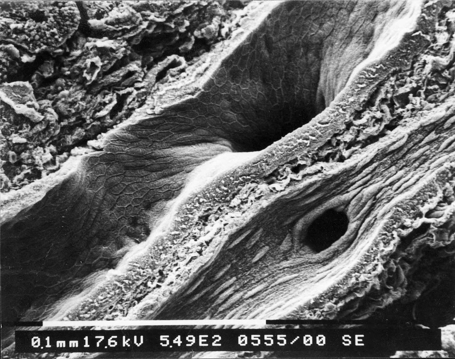
Salivary duct and artery in a salivary gland
This is an electron microscopic image taken of a tissue sample from a salivary gland. There are lots of interesting structures to be seen.
Crossing the center of the image is a salivary duct (terminal excretory duct). The borders between the epithelial cells lining the inner surface of the salivary duct can be seen on its inner surface. There is also a branch of the salivary duct that runs into the tissue displaying a hole in the wall of the duct.
Running alongside the salivary duct is an artery. It can be identified by its many protuberances lining its inner surface. These "bumps" are actually the nuclei of the smooth muscle cells of the artery. The artery also displays branching of a smaller vessel in its wall (black hole in the wall of the artery).
At the top left, an acinus is cut open.