


Radix of the tongue (40X)
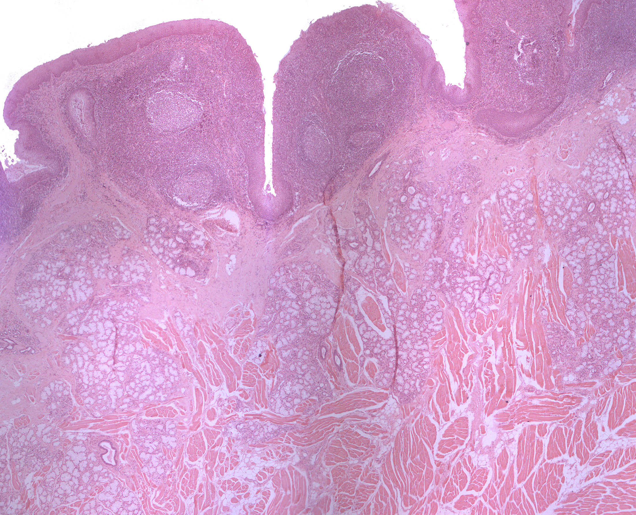
This frontal section is taken from the posterior part of the tongue , the lingual radix (lat: lingua). Stratified squamous epithelium covers the lamina propria. You can see lymphoid tissue, the lingual tonsils, some adipose cells, numerous vascular beds and a few nerves. Within the deeper parts of the lamina propria and the connective tissue, you can find quite a few mucous acini. Their excretory ducts open on the dorsum (upper surface) of the tongue.